industryterm:medical imaging

  • A beginner’s guide to Deep Learning Applications in Medical Imaging
    https://hackernoon.com/a-beginners-guide-to-deep-learning-applications-in-medical-imaging-7aa3b

    Let us first understand what medical imaging is before we delve into how deep learning and other similar expert systems can help medical professional such as radiologists in diagnosing their patients.This is how Wikipedia defines Medical Imaging:Medical imaging is the technique and process of creating visual representations of the interior of a body for clinical analysis and medical intervention, as well as visual representation of the function of some organs or tissues (physiology). Medical imaging seeks to reveal internal structures hidden by the skin and bones, as well as to diagnose and treat disease. Medical imaging also establishes a database of normal anatomy and physiology to make it possible to identify abnormalities. Although imaging of removed organs and tissues can be (...)

    #keras #deep-learning #artificial-intelligence #medicine #machine-learning

  • CppCast Episode 166: CppCon Poster Program and Interface Design with Bob Steagall
    http://isocpp.org/feeder/?FeederAction=clicked&feed=All+Posts&seed=http%3A%2F%2Fisocpp.org%2Fblog%2F2

    Episode 166 of CppCast the only podcast for C++ developers by C++ developers. In this episode Rob and Jason are joined by Bob Steagall to discuss his history with C++, the CppCon poster program and his upcoming talks.

    CppCast Episode 166: CppCon Poster Program and Interface Design with Bob Steagall by Rob Irving and Jason Turner

    About the interviewee:

    Bob is a Principal Engineer with GliaCell Technologies. He’s been working almost exclusively in C++ since discovering the second edition of The C++ Programming Language in a college bookstore in 1992. The majority of his career was spent in medical imaging, where he led teams building applications for functional MRI and CT-based cardiac visualization. After a brief detour through the worlds of DNS and analytics, he’s (...)

    #News,Video&_On-Demand,

  • Facebook and NYU School of Medicine launch research collaboration to improve MRI – Facebook Code
    https://code.fb.com/ai-research/facebook-and-nyu-school-of-medicine-launch-research-collaboration-to-improv

    C’est bô le langage fleuri des experts en public relation...

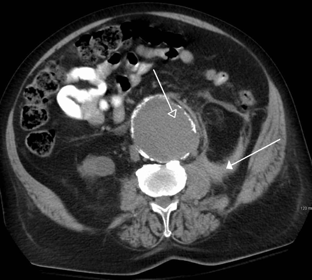
    Using AI, it may be possible to capture less data and therefore scan faster, while preserving or even enhancing the rich information content of magnetic resonance images. The key is to train artificial neural networks to recognize the underlying structure of the images in order to fill in views omitted from the accelerated scan. This approach is similar to how humans process sensory information. When we experience the world, our brains often receive an incomplete picture — as in the case of obscured or dimly lit objects — that we need to turn into actionable information. Early work performed at NYU School of Medicine shows that artificial neural networks can accomplish a similar task, generating high-quality images from far less data than was previously thought to be necessary.

    In practice, reconstructing images from partial information poses an exceedingly hard problem. Neural networks must be able to effectively bridge the gaps in scanning data without sacrificing accuracy. A few missing or incorrectly modeled pixels could mean the difference between an all-clear scan and one in which radiologists find a torn ligament or a possible tumor. Conversely, capturing previously inaccessible information in an image can quite literally save lives.

    Advancing the AI and medical communities
    Unlike other AI-related projects, which use medical images as a starting point and then attempt to derive anatomical or diagnostic information from them (in emulation of human observers), this collaboration focuses on applying the strengths of machine learning to reconstruct the most high-value images in entirely new ways. With the goal of radically changing the way medical images are acquired in the first place, our aim is not simply enhanced data mining with AI, but rather the generation of fundamentally new capabilities for medical visualization to benefit human health.

    In the interest of advancing the state of the art in medical imaging as quickly as possible, we plan to open-source this work to allow the wider research community to build on our developments. As the project progresses, Facebook will share the AI models, baselines, and evaluation metrics associated with this research, and NYU School of Medicine will open-source the image data set. This will help ensure the work’s reproducibility and accelerate adoption of resulting methods in clinical practice.

    What’s next
    Though this project will initially focus on MRI technology, its long-term impact could extend to many other medical imaging applications. For example, the improvements afforded by AI have the potential to revolutionize CT scans as well. Advanced image reconstruction might enable ultra-low-dose CT scans suitable for vulnerable populations, such as pediatric patients. Such improvements would not only help transform the experience and effectiveness of medical imaging, but they’d also help equalize access to an indispensable element of medical care.

    We believe the fastMRI project will demonstrate how domain-specific experts from different fields and industries can work together to produce the kind of open research that will make a far-reaching and lasting positive impact in the world.

    #Resonance_magnetique #Intelligence_artificielle #Facebook #Neuromarketing

  • Data Visualization, Design and Information Munging // Martin Krzywinski / Genome Sciences Center

    http://mkweb.bcgsc.ca

    With some very smart people, I work on problems in data visualization applied to cancer research and genome analysis. Previously I was involved in fingerprint mapping, system administration, computer security, fashion photography, medical imaging and LHC particle physics. My work is guided by a need to rationalize, make things pretty, combine science with art, mince words, find good questions and help make connections between ideas. All while exercising snark.

    #visualisation #solutions #graphiques #cartographie

  • A shape model – what’s that?
    http://blogs.esa.int/rosetta/2015/08/13/a-shape-model-whats-that

    Today, a new shape model of Rosetta’s comet is released by ESA. Some of you might immediately know what this is and how you might use it, and others will wonder ‘what’s that?’ This blog post, which has been prepared with the help of experts from the Rosetta Flight Dynamics team, explains what a shape model is, how it is created, and what you could use it for. At the most basic level, a shape model is a geometrical representation of an object. Shape models are commonly used in computer programmes where the motion or change of shape of a complex object needs to be represented. Applications can vary from medical imaging of organs, creating characters in cartoons and computer games, or — closer to home (at least for the Rosetta team) — modelling how the surface of a comet changes as it rotates. (...)

  • Metals Used in High Tech Are Becoming Harder to Find, Study Says
    http://e360.yale.edu/digest/metals_used_in_high_tech_are_becoming_harder_to_find_study_says/4398
    http://environment.yale.edu/content/images/00006518/yale-criticality-metals-3d-graphic-browser.jpg?1427467310

    Metals critical to newer technologies such as #smartphones, infrared optics, and medical imaging will likely become harder to obtain in coming decades, according to Yale researchers, and future products need to be designed to make reclaiming and recycling those materials easier.
    http://environment.yale.edu/news/article/metals-used-in-hightech-products-face-future-supply-risks

    The study, the first to assess future supply risks to all 62 metals on the periodic table, found that many of the metals traditionally used in manufacturing — zinc, copper, aluminum, lead, and others — show no signs of vulnerability. But some metals that have become more common in technology over the last two decades, such as rare earth metals, are available almost entirely as byproducts of other elements, the researchers say.
    http://e360.yale.edu/feature/a_scarcity_of_rare_metals_is_hindering_green_technologies/2711

    "You can’t mine specifically for them; they often exist in small quantities and are used for specialty purposes," said Yale scientist Thomas Graedel. "And they don’t have any decent substitutes." Metals such as lead are highly recycled because they’re often used in bulk. But recycling the relatively rare metals critical to modern electronics is far more difficult because they are used in miniscule amounts and can be difficult to extract from complex and compact new technologies, Graedel said.

    #métaux_rares #écrans #DEEE

  • We Are Giving Ourselves #Cancer
    http://www.nytimes.com/2014/01/31/opinion/we-are-giving-ourselves-cancer.html?_r=0

    DESPITE great strides in prevention and treatment, cancer rates remain stubbornly high and may soon surpass heart disease as the leading cause of death in the United States. Increasingly, we and many other experts believe that an important culprit may be our own medical practices: We are silently irradiating ourselves to death.

    The use of medical imaging with high-dose radiation — CT scans in particular — has soared in the last 20 years. Our resulting exposure to medical radiation has increased more than sixfold between the 1980s and 2006, according to the National Council on Radiation Protection & Measurements. The radiation doses of CT scans (a series of X-ray images from multiple angles) are 100 to 1,000 times higher than conventional X-rays.

    (...)

    The relationship between radiation and the development of cancer is well understood: A single CT scan exposes a patient to the amount of radiation that epidemiologic evidence shows can be cancer-causing. The risks have been demonstrated directly in two large clinical studies in Britain and Australia. In the British study, children exposed to multiple CT scans were found to be three times more likely to develop leukemia and brain cancer. In a 2011 report sponsored by Susan G. Komen, the Institute of Medicine concluded that radiation from medical imaging, and hormone therapy, the use of which has substantially declined in the last decade, were the leading environmental causes of breast cancer, and advised that women reduce their exposure to unnecessary CT scans.

  • Vast Effort by FDA Spied on E-Mails of Its Own Scientists
    http://readersupportednews.org/news-section2/318-66/12421-vast-effort-by-fda-spied-on-e-mails-of-its-own-scientists

    The agency, using so-called spy software designed to help employers monitor workers, captured screen images from the government laptops of the five scientists as they were being used at work or at home. The software tracked their keystrokes, intercepted their personal e-mails, copied the documents on their personal thumb drives and even followed their messages line by line as they were being drafted, the documents show.

    The extraordinary #surveillance effort grew out of a bitter dispute lasting years between the scientists and their bosses at the F.D.A. over the scientists’ claims that faulty review procedures at the agency had led to the approval of medical imaging devices for mammograms and colonoscopies that exposed patients to dangerous levels of radiation.

    #santé #whistle-blowers via @opironet